AHA PALS and ScvO2 Monitoring
PALS has changed drastically in the last ten years, and technology and research are leading that change. Queue continuous central venous oxygen monitoring, a hallmark of the 2020/2025 PALS algorithms.
Understanding ScvO2 requires a bit of understanding of physiology. Remember that, as hemoglobin molecules leave the heart after being oxygenated in the lungs, they have four molecules of oxygen attached. Out in the tissue of the body, one oxygen molecule is extracted, leaving the RBCs 75% saturated as they return through the venae cavae to the heart. Measurement of the level of saturation of RBCs as they return to the heart (venous oxygenation) has been done for some time, but what's new is the continuous measurement of levels.
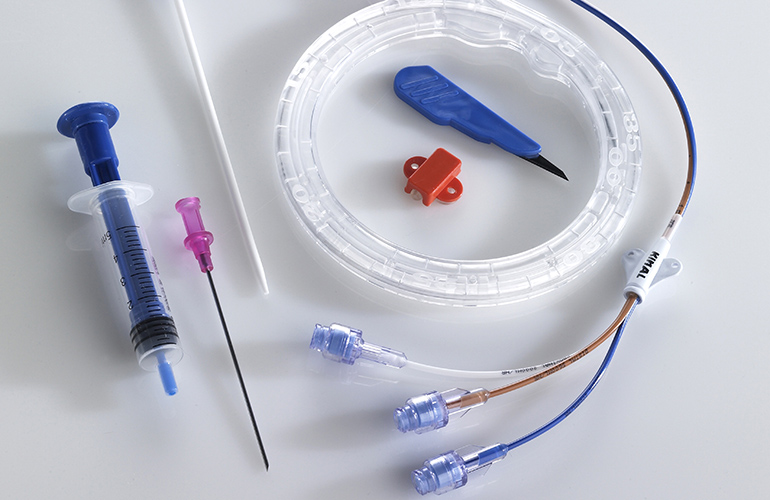
Continuous venous oxygen monitoring can be performed in two locations—the superior vena cava (venous return from the upper body) or the pulmonary artery (which contains venous blood from the upper body, lower body, and coronary veins). When the catheter is placed in the SVC, we call the resulting value "ScvO2" (saturation of central venous oxygen). When the catheter is placed in the PA, we call the value "SvO2" (venous oxygen saturation). PALS algorithms utilize ScvO2. ScvO2 levels are always 5 to 10% higher than SvO2 levels, but thankfully for us, they correlate exactly.
Why does AHA use ScvO2 instead of SvO2? Most critically ill patients will require the use of a central line anyway, and a triple lumen central venous catheter with fiber optic ScvO2 measurement can be placed, saving the patient from more invasive PA line placement. The most accurate ScvO2 catheters use three wavelengths of light, which can measure oxygenation independent of hemoglobin levels. All that being said, the obvious question that remains is, "Why do I care about ScvO2? I have other parameters at hand."
The answer is both simple and profound. ScvO2 gives you an indication of oxygenation problems earlier than any other parameter. To spell this out, continuous ScvO2 monitoring can tell you about increased oxygen consumption (due to infection or hyperthermia), decreased cardiac output (due to hypovolemia or sepsis), or decreased oxygen supply (due to respiratory failure) first. The normal range of ScvO2 is 70–80%. When values drop by 10% or below 70%, intervention is indicated. The patient still compensates for decreased oxygenation in the 50–70% ScvO2 range, at which point other parameters may still not show warning signs. By the time other parameters (like SpO2) do, damage to tissues is already occurring.
Fundamental PALS concepts can be learned through an AHA PALS course. Find the best AHA PALS course in Austin, Texas at Thri Austin - an official local Austin AHA Training Site offering BLS, PALS, and ACLS. While Thri Austin offers same-day AHA certifications with its RQI training system, the Thri network of official AHA Training Sites also features non-RQI AHA Training like Los Angeles Heart Training, which offers official AHA PALS classes in LA and also AHA BLS and AHA ACLS in Los Angeles.
Author
CHT Staff
At Chicago Heart Training we believe active medical practice should be supported by a foundation of rigorous research in medicine and health care practice. Our staff engages in regular investigation of trending issues in medicine and provides research-backed opinion articles to enrich your medical tastes.